Molybdenum ditelluride (MoTe2) belongs to a large group of materials with a layered structure, also known as 2D materials. This means that their properties (e.g. electric) change significantly when the material is extremely thin, consisting of only a few atomic planes stacked on top of each other. This property distinguishes layered materials from bulk materials.
MoTe2 occurs in several different crystal structures. Each of these structures has different electronic properties: the hexagonal MoTe2 is a semiconductor, the monoclinic is a semimetal, and the orthorhombic structure is a metal. These properties can be affected by various external influences, such as changes in temperature, pressure, and external electric field. Therefore, these materials (and thin layers prepared from them) can be used in electronics as sensors, detectors, memories, or even in solar cells. For these reasons, it is useful to know the detailed crystalline structure of the thin films and how they are arranged after deposition on the substrate.
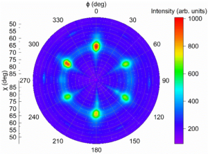
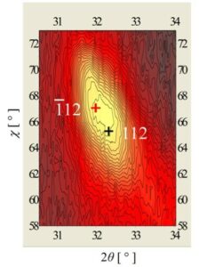
Our colleagues recently published work where they studied thin layers of MoTe2 with a thickness of 10-40 nm on a sapphire substrate with two different structures – hexagonal and monoclinic. We examined these layers using X-ray diffraction (XRD) analysis. The XRD diffraction pattern is characteristic for particular material and enables not only description but also complete disclosure of its structure. In the analysis, we used 2 different complementary X-ray analysis techniques: standard XRD and wide-angle scattering at a small angle of incidence (GIWAXS – Grazing Incidence Wide Angle X-Ray Scattering). Standard XRD provides accurate values of the lattice parameters and orientation of the sample on the substrate. The GIWAX technique gives an overall picture of the available diffractions in the reciprocal space because it uses an area detector. It was found that the sapphire substrate “imposed” its symmetry on the MoTe2 layers, as shown by the so-called pole figures whose symmetry reflects the symmetry of the material (Fig. 1). The analysis of the structure can also be complicated by the overlapping of close but not the same diffractions originating from differently rotated single crystal domains in the thin layer. Such diffractions exist due to the presence of six sets of single crystal domains rotated by multiples of 60° and, due to insufficient angular resolution, difficult to distinguish in the GIWAXS pattern (Fig. 1). The solution provided a high-resolution diffraction measurement. In a special, so-called 2θ/χ map (Fig. 2), it is possible to recognize the presence of two diffractions, which confirms the formation of the monoclinic MoTe2 phase.
Pribusová Slušná, L., Vegso, K., Dobročka, E., Vojteková, T., Nádaždy, P., Halahovets, Y., Sojková, M., Hrdá, J., Precner, M., Šiffalovič, P., Chen, Z., Huang, Y., Ražnjević, S., Zhang, Z., and Hulman, M.: Ordered growth of hexagonal and monoclinic phases of MoTe2 on a sapphire substrate, CrystEngComm 25 (2023) 5706-5713.