Magnetic force microscopy (MFM) has become established as a standard method to probe surface magnetic textures of a large variety of magnetic materials.
Working principle of MFM is based on an oscillating cantilever ended by a sharp tip covered by magnetic material. During the scanning, this magnetic tip is passed closely above the sample surface and the magnetostatic forces between the tip and the sample affect the phase and frequency of cantilever oscillations, which enables to detect magnetic profile of the sample surface. Reducing the diameter of the tip apex (i.e. increasing its sharpness) the resolution of the MFM can be improved up to the 10 nm level. However, such super sharp tips are prone to be deformed when the tip impacts the sample surface. As a result, their magnetic moment changes with time and the tips can be even destroyed, which hinders their desired utility in quantitative MFM scanning providing the distribution of magnetic charges at the sample surface.
In our previous experimental work, we successfully demonstrated a new type of MFM probe with a ferromagnetic circular disk at its apex. The magnetic structure of the disk is in a vortex state, i.e. most of its magnetic moments circulate in-plane around the disk center and generate zero magnetic field. Only a small fraction of spins located in the vicinity of the disc center (dubbed as a vortex core) is oriented out of the disk plane and serves as a super sharp magnetic probe. Since the disk diameter is relatively large, our vortex-core probe is robust against mechanical deformations, and, at the same time, its magnetic properties (e.g. resolution) are similar to the commercial super sharp tips.
However, the ferromagnetic disk is made of soft magnetic material (Permalloy) which is sensitive to the larger magnetic field from the sample and its magnetization can be undesirably altered during the scanning process. Therefore, in the present work, we study numerically the limitations of the vortex-core probe by calculating the scanned images of typical samples, i.e. isolated circular domains, magnetic stripes, and domains in chessboard arrangement mimicking the bits in computer hard drives. Our results show that the vortex core probe provides a reliable MFM image when scanning the flat samples with magnetic domains sized up to 100 nm. For larger domains, the magnetic field they generate is strong enough to introduce unwanted artifacts into the resulting MFM scan.
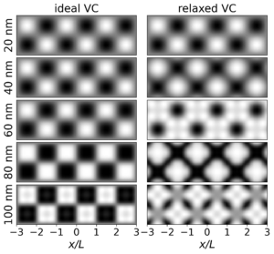 Left panel: Simulated MFM image of square domains with various sizes L made by ideal VC probe. Right: Simulated MFM image of the same samples made by the VC probe with magnetization deformed by the magnetic field of the sample.
Feilhauer, J., Tóbik, J., Šoltýs, J., and Cambel, V.: Numerical characterization of magnetic vortex probe imaging for magnetic force microscopy, IEEE Trans. Magnet. 59 (2023) 6500210.
Left panel: Simulated MFM image of square domains with various sizes L made by ideal VC probe. Right: Simulated MFM image of the same samples made by the VC probe with magnetization deformed by the magnetic field of the sample.
Feilhauer, J., Tóbik, J., Šoltýs, J., and Cambel, V.: Numerical characterization of magnetic vortex probe imaging for magnetic force microscopy, IEEE Trans. Magnet. 59 (2023) 6500210.